Cardiovascular Magnetic Resonance Imaging (CMR)
Heart Anatomy
The heart is considered to have three segments: the atria, ventricles and the great vessels.
The atria and ventricles are each partitioned into two counterparts, the left and right sides, with two atrioventricular valves (tricuspid valve and mitral valve) that connect the respective ventricle to the atria and the semilunar valves (pulmonary valve, aortic valve) that connect the great vessels to the ventricles.
On the other hand, the right atrium (green chamber in Fig. 1c) (see Fig. 1b). The right ventricle (blue chamber) (brown artery in Fig. 1c).
The left atrium (LA) receives oxygenated blood via the pulmonary veins and is connected to the left ventricle through the mitral valve which has two leaflets
The left ventricle (LV) is the main pumping chamber that pumps blood through the aorta causing the three leaflets aortic valve (see Fig. 1b) to open during systole and to close during diastole.
The right atrium (RA) receives deoxygenated blood from the superior and inferior vena cava and is connected to the right ventricle through the tricuspid valve that has three leaflets.
The right ventricle (RV) pumps blood through the three-leaflets pulmonary valve towards the pulmonary artery.
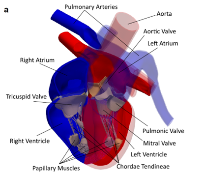 The blue part corresponds to deoxygenated blood, while the red part corresponds to oxygenated blood

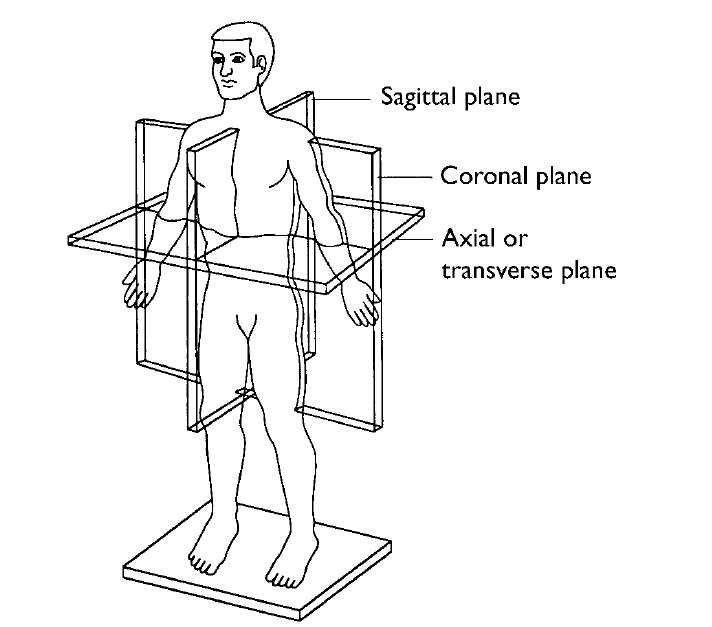
Imaging Planes of the Body
The body is divided into three planes:
- The axial plane (transverse, Z) plane that cuts the body from top to bottom (or superior to inferior).
- The coronal plane (Y) plane that cuts the body from front to back (or anterior to posterior).
- The sagittal plane (X) plane that cuts the body from right to left.
Overview of CMR Protocol for UK Biobank
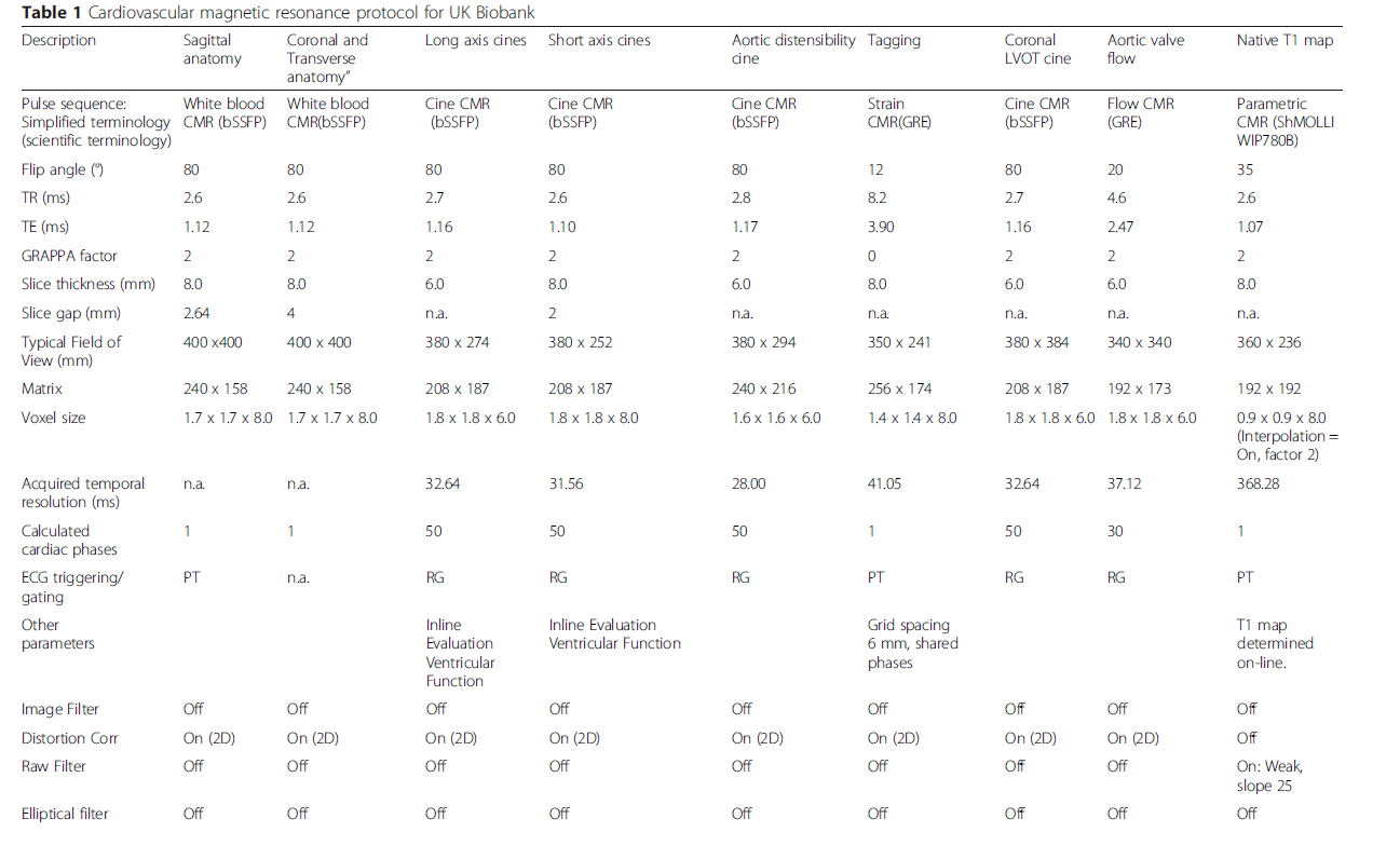 (Petersen et al., 2015)
The information for each modality will be detailed in each section.
Reference Ranges of Features
For each feature, the corresponding reference range can be found in /tasks/reference_range.json file.
Most features can be indexed using BSA, calculated using the DuBois & DuBois formula: , where is the height in cm and is the weight in kg (Du Bois & Du Bois, 1916).
TODO:
- Number of cusps of aortic valve
- 3Ch View
- Enhancement of registration for strain
- Changes in angle caused by shear (ERC, ECL)
- More strain calculated from tagged MRI
References
M.D, R. W. B., Biederman, R. W. W., Doyle, M., & Yamrozik, J. (2008). The Cardiovascular MRI Tutorial: Lectures and Learning. Lippincott Williams & Wilkins.
Viola, F., Del Corso, G., De Paulis, R., & Verzicco, R. (2023). GPU accelerated digital twins of the human heart open new routes for cardiovascular research. Scientific Reports, 13(1), 8230. https://doi.org/10.1038/s41598-023-34098-8
Petersen, S. E., Matthews, P. M., Francis, J. M., Robson, M. D., Zemrak, F., Boubertakh, R., Young, A. A., Hudson, S., Weale, P., Garratt, S., Collins, R., Piechnik, S., & Neubauer, S. (2015). UK Biobank’s cardiovascular magnetic resonance protocol. Journal of Cardiovascular Magnetic Resonance, 18(1), 8. https://doi.org/10.1186/s12968-016-0227-4
Du Bois D, Du Bois EF. A formula to estimate the approximate surface area if height and weight be known. 1916. Nutrition 1989;5:303311; discussion 312–303.