Evaluation of Atrial Volume
The LA volumes are derived using the biplane area-length method, which is based on the formula (Nacif et al., 2012).
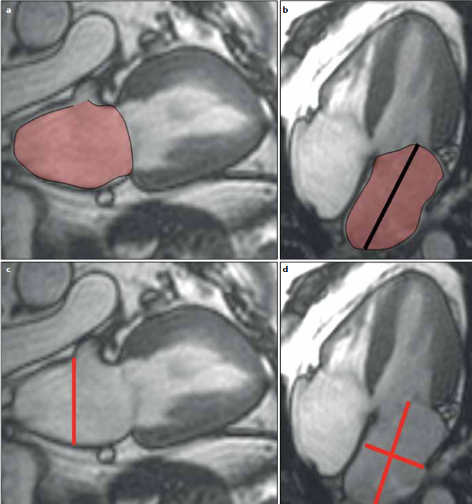
Nacif, M. S., Barranhas, A. D., Türkbey, E., Marchiori, E., Kawel, N., Mello, R. A. F., Falcão, R. O., Junior, A. C. O., & Rochitte, C. E. (2012). Left atrial volume quantification using cardiac MRI in atrial fibrillation: Comparison of the Simpson’s method with biplane area-length, ellipse, and three-dimensional methods. Diagnostic and Interventional Radiology. https://doi.org/10.5152/dir.2012.002
Sphericity index was calculated as the ratio of LA maximum volume to the volume of a sphere with maximum LA length diameter among atrial length and transverse length
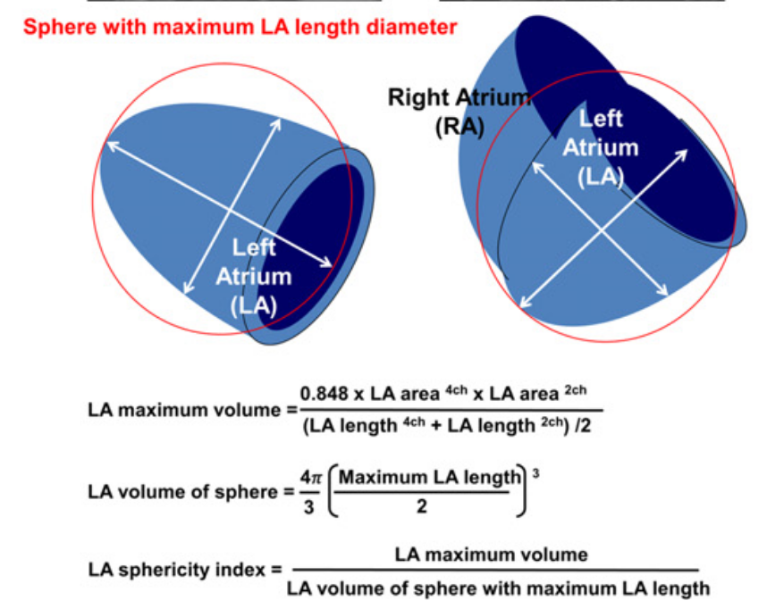
Aquaro, G. D., Pizzino, F., Terrizzi, A., Carerj, S., Khandheria, B. K., & Di Bella, G. (2019). Diastolic dysfunction evaluated by cardiac magnetic resonance: The value of the combined assessment of atrial and ventricular function. European Radiology, 29(3), 1555–1564. https://doi.org/10.1007/s00330-018-5571-3
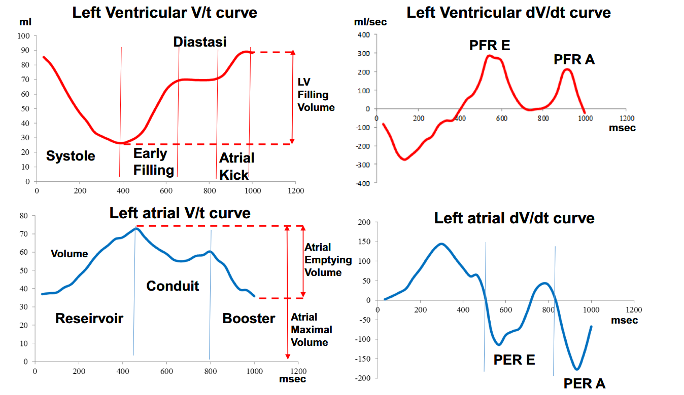
After deriving the atrial and ventricular dV/dt curves from the volume curves. Peaks of the ventricular dV/dt curves were defined as follows: the first positive peak was defined as the early peak filling rate (PFR-E) and the second peak was defined as atrial peak filling rate (PFR-A). PFR-E and PFR-A represent the maximum speed of passive filling and the maximum speed of filling secondary to atrial contraction, respectively. Between the two peaks, a zone of almost zero speed is normally present; this is the phase of diastasis.
Nakamori, S., Ngo, L. H., Tugal, D., Manning, W. J., & Nezafat, R. (2018). Incremental Value of Left Atrial Geometric Remodeling in Predicting Late Atrial Fibrillation Recurrence After Pulmonary Vein Isolation: A Cardiovascular Magnetic Resonance Study. Journal of the American Heart Association, 7(19), e009793. https://doi.org/10.1161/JAHA.118.009793
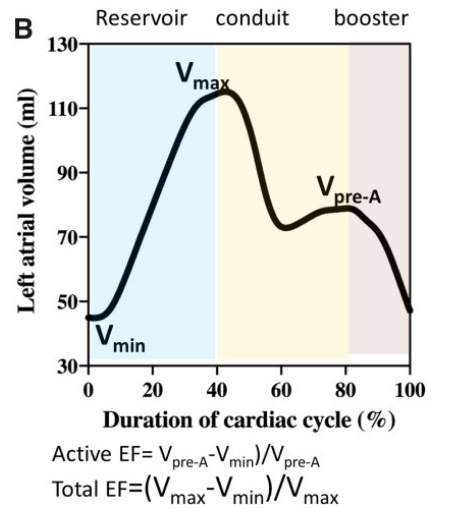
Left atrial function has been typically divided into three integrated phases: reservoir, conduit and booster-pump (Figure 1) [7–10]. Reservoir: an expansion phase during left ventricular (LV) systole; the LA stores pulmonary venous return during LV contraction and isovolumic relaxation. Conduit: the LA transfers blood passively into the LV during ventricular diastole. Booster-pump: contractile component (when supraventricular rhythm is present);
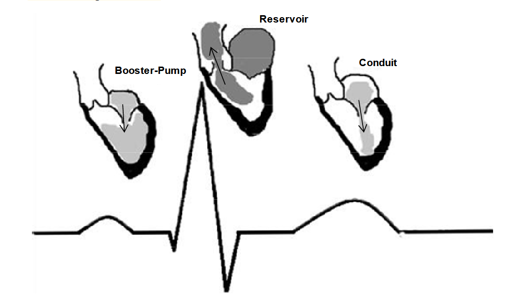
Mehrzad, R., Rajab, M., & Spodick, D. (2014). The Three Integrated Phases of Left Atrial Macrophysiology and Their Interactions. International Journal of Molecular Sciences, 15(9), 15146–15160. https://doi.org/10.3390/ijms150915146
Peters, D. C., Lamy, J., Sinusas, A. J., & Baldassarre, L. A. (2021). Left atrial evaluation by cardiovascular magnetic resonance: Sensitive and unique biomarkers. European Heart Journal Cardiovascular Imaging, 23(1), 14–30. https://doi.org/10.1093/ehjci/jeab221

Hoit, B. D. (2014). Left Atrial Size and Function. Journal of the American College of Cardiology, 63(6), 493–505. https://doi.org/10.1016/j.jacc.2013.10.055